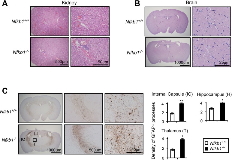
Figure 2. Nfkb1−/− mice exhibit enhanced age-related CNS gliosis.
Representative H&E stained kidney (A) and brain (B) sections from 12-month old Nfkb1+/+ and Nfkb1−/− mice (n=4 per group). (C) Representative brain sections (left) from 12-month old Nfkb1+/+ and Nfkb1−/− mice stained with anti-GFAP antibody. IC-internal capsule, H-hippocampus, T-thalamus. Graphs (right) demonstrate density of GFAP processes within the indicated region determined by averaging the level of staining from three separate fields in four coronal sections, separated by 3 mm (A-P diameter). n=4 animals per group. Scale: 1- very few GFAP processes; 2: <25% staining; 3: 25-50% staining and 4: >50% staining. Data represent mean +/− SEM. *p<0.03. **p<0.01.

