Fig. 3.
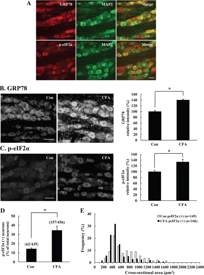
Immunofluorescence analysis of GRP78 and p-eIF2α in CFA-injected rat TG. On the fifth day after CFA-injection in rat vibrissal pad, we examined the expression of ER stress marker proteins in rat TG. (A) In control rats, GRP78 and p-eIF2α were mostly expressed in TG neurons, as demonstrated by colocalization of GRP78 or p-eIF2α (red) and neuronal marker MAP2 (green). (B-C) GRP78 or p-eIF2α immunoreactivity increased remarkably in CFA-injected TG compared to the control TG. Quantification of the GRP78 or p-eIF2α immunopositive intensity increased in CFA-injected TG compared to control TG (right). (D) A significant increase in the number of p-eIF2α immunopositive neurons was observed in the CFA-injected TG. (E) The expression of p-eIF2α increased significantly in small and medium sized neurons after CFA-induced peripheral inflammation. Data is expressed as means±SD of three independent experiments. Scale bar=50 µm. *p<0.05 (Student's t-test).

