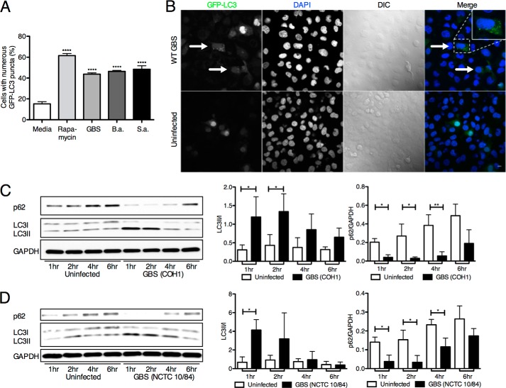
FIGURE 1.
Autophagy induction in hBMECs. A, GFP-LC3 (Ad-GFP-LC3) counts in hBMECs following infection with GBS (COH1 WT), B. anthracis (B.a.) (Sterne 7702 WT), and S. aureus (S.a.) (ISP479C WT) for 3 h (m.o.i. = 10) compared with untreated controls. At least 200 cells with numerous puncta were counted, and rapamycin treatment (5 μm) was used as a positive control. Significance was measured in comparison with untreated controls. B, confocal microscopy visualization of COH1 WT and uninfected hBMECs transduced with GFP-LC3. Arrows denote cells with abundant amounts of puncta. Scale bar, 10 μm. C and D, Western blot analysis of LC3 and p62 in hBMEC samples at various time points postinfection with COH1 for 4 h (m.o.i. = 10) and NCTC 10/84 for 1 h (m.o.i. = 10). Image analysis was performed using ImageJ software to determine LC3-II/LC3-I and p62/GAPDH ratios. All experiments were repeated at least three times in triplicate; data represent the mean ± S.D. from a representative experiment. *, p < 0.05; **, p < 0.005; ****, p < 0.0001. Error bars represent S.D. DIC, differential interference contrast.

