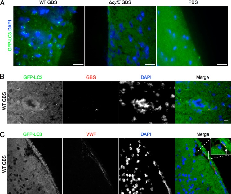
FIGURE 6.
Visualization of autophagy activation in brain endothelium. A, representative brain samples from GFP-LC3 transgenic mice infected with NCTC 10/84 GBS and isogenic ΔcylE mutant. GFP-LC3 puncta were observed in brain tissue in WT-infected mice compared with mice infected with the ΔcylE mutant or injected with PBS. Scale bar, 20 μm. B, immunofluorescence for GBS in WT-infected GFP-LC3 transgenic mice. GBS co-localizes with GFP-LC3 within endothelial portions of the brain. Scale bar, 10 μm. C, immunofluorescence for von Willebrand factor (VWF) in GFP-LC3 transgenic WT-infected mice demonstrates that endothelial cells are producing active GFP-LC3. Scale bar, 10 μm.

