FIGURE 8.
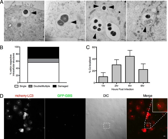
Examination of the intracellular localization and LC3 co-localization of GBS. A and B, transmission electron microscopy of intracellular GBS COH1 4 h after hBMEC infection (m.o.i. = 10). Intracellular bacteria were quantified according to the intracellular structure in which they resided (n = 25). Bacteria are present in membrane-bound vesicles, damaged membranes, multiple membranes, and putative autophagic structures as indicated by arrowheads. Scale bar, 500 nm. C, transfection of an mCherry-LC3 plasmid into hBMECs was performed as described under “Experimental Procedures.” hBMECs were infected with GFP COH1 WT for 4 h prior to treatment with extracellular antibiotics 1, 2, 4, and 6 h postinfection. Quantification of the amount of GFP COH1 WT co-localizing with mCherry-LC3 was gathered by counting at least 100 cells with intracellular GBS, and data are means ± S.E. from a representative experiment performed in triplicate. D, representative confocal microscopic images from 4 h postinfection. Scale bar, 10 μm. Error bars represent S.D. DIC, differential interference contrast.

