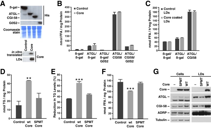
FIGURE 5.
Core and ATGL association with LDs is necessary to inhibit lipolysis. A, Western blot of Cos-7 cell lysates expressing β-gal, ATGL, CGI-58, or G0S2, in vitro translated core, or isolated LD fractions from Cos-7 cells expressing core or an empty vector. B, cell lysates of Cos-7 cells overexpressing ATGL, CGI-58, G0S2, or β-gal as control were incubated with a radiolabeled triolein substrate in the presence or absence of in vitro translated core protein for 1 h at 37 °C. FFAs were then extracted, and radioactivity was determined by liquid scintillation counting. Results are representative of two independent experiments with three replicates in each experiment. Data are presented as the mean ± S.D. C, radiolabeled LDs were isolated from Cos-7 cells transfected with core or an empty vector and then incubated with Cos-7 cell lysates expressing ATGL, CGI-58, and G0S2 or β-gal as the control for 1 h at 37 °C. FFAs were then extracted, and radioactivity was determined by liquid scintillation counting. Results are representative of two independent experiments with three replicates in each experiment. Data are presented as the mean ± S.D. D–F, NIH/3T3 were transduced with a lentivirus encoding HA-core WT or SPMT mutant and GFP or a control virus encoding GFP alone. D, 24 h after infection, neutral lipids were extracted, and TGs were measured and normalized to total protein. E and F, cells were incubated with oleic acid for 20 h before a 6-h chase using medium with 2% fatty acid-free BSA and 10 μm triacsin C. Neutral lipids were extracted, the amount of TG was measured and normalized to T0 (E). FFA levels were measured in the culture supernatant (F). Both TG and FFA levels were normalized to the amount of cellular protein. Results are representative of three independent experiments with three replicates in each experiment. Data are presented as the mean ± S.D. G, Western blot of cell extracts (left panels) or isolated LD fractions (right panels) from Huh7-Lunet cells expressing core WT or SPMT mutant. Results are representative of two independent experiments.

