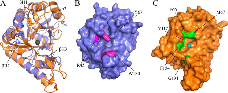
FIGURE 4.

IcaBAd has a narrow active site pocket compared with PgaB43–309. A, superposition of IcaBAd23–280Δloop (blue) and PgaB43–309 (orange) shows a strong conservation of the CE4 fold with a root mean square deviation of 1.7 Å over 219 equivalent Cα atoms. β-Hairpin loops for IcaBAd are labeled βH1–3, and the Zn2+ ion is shown as a sphere colored dark gray. Surface representation of IcaBAd23–280Δloop (B) and PgaB43–309 (C) is shown in the same representation as panel A with residues contributing to the difference in active site architecture colored magenta and green for IcaBAd23–280Δloop and PgaB43–309, respectively. The Ni2+ ion in PgaB is shown as a teal-colored sphere.
