
FIGURE 7.
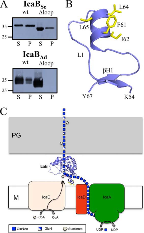
Membrane localization of IcaB. A, staphylococcal membrane pulldown assays of purified IcaB proteins show the hydrophobic loop is important for membrane association. S, supernatant; P, pellet; wt, wild type. Molecular mass markers are indicated on the left side of the blots in kDa. B, predicted structure for the IcaBSe hydrophobic loop reveals an amphipathic α-helix. C, proposed model for PNAG biosynthesis and modification. IcaB associates with the membrane via its hydrophobic loop and electropositive surface and de-N-acetylates PNAG after export. M, membrane; PG, peptidoglycan; CoA, coenzyme A.