
FIGURE 1.
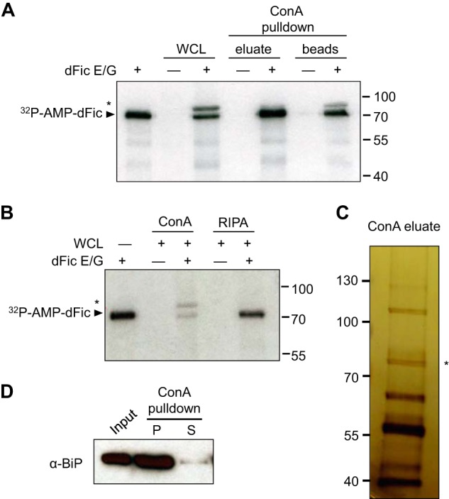
BiP is identified as a substrate for dFic. A, recombinant GST-dFicΔ70 E247G (dFic E/G) was incubated with S2 whole cell lysate and the ConA-bound fraction of lysate in the presence of α-32P-labeled ATP. The arrowhead indicates the autoAMPylated dFic, and the asterisk marks the putative substrate of the ∼72-kDa from the lysate labeled with dFic. WCL, whole cell lysate. B, GST-dFicΔ70 E247G was incubated with S2 whole cell lysate prepared with either standard radioimmune precipitation assay buffer (RIPA) or RIPA buffer added with 5 mm of Mg2+, Mn2+, and Ca2+ (ConA), which is required for concanavalin A binding. C, concanavalin A-bound proteins were eluted from the beads, concentrated, and analyzed by silver stain after SDS-PAGE. The asterisk indicates a single band with the size corresponding to the putative substrate of dFic observed from previous AMPylation assays. D, S2 cell lysate was incubated with ConA-agarose, and the bound (P) and unbound (S) proteins were analyzed by anti-BiP.