FIGURE 3.
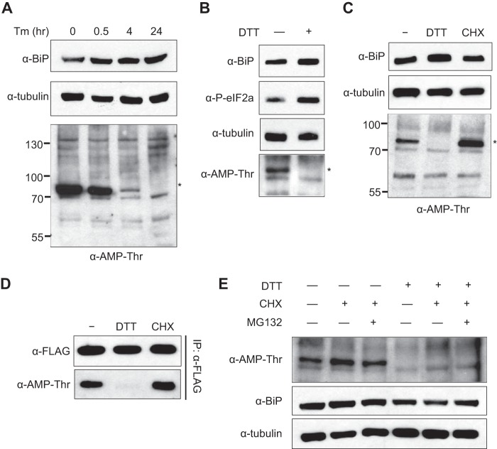
AMPylation of BiP is modulated by ER stress. A, pharmacological ER stress inducer tunicamycin (Tm) was treated to S2 cells for the indicated times at 2 μg/ml. Whole cell lysates from different time points were analyzed by anti-BiP and anti-Thr-AMP. Anti-tubulin was used as a loading control. The asterisk marks the AMPylated protein from cell lysate with ∼72 kDa. B, 5 mm DTT was used to trigger ER stress in S2 cells for 1 h, and the whole cell lysates were analyzed with the indicated antibodies. Anti-phospho-eIF2α was used as a marker for ER stress. C, 5 mm DTT and 100 μg/ml cycloheximide (CHX), a potent inhibitor of protein synthesis, was added to S2 cells for 4 h. Whole cell lysates were analyzed with indicated antibodies. D, cells were transfected with BiP containing a C-terminal FLAG tag followed by DTT and cycloheximide treatment for 4 h. BiP was then immunoprecipitated with anti-FLAG-agarose and analyzed with anti-BiP and anti-Thr-AMP. Immunoprecipitation was confirmed by anti-FLAG. E, cells were treated with 5 mm DTT and/or 100 μg/ml cycloheximide in the presence or absence of 20 μm MG132 for 4 h.

