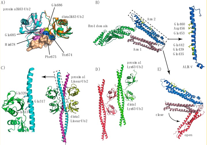
Figure I.
(A) Different X-ray structures of the TAB2NZF/Lys63-Ub2 complex, PDB code 3A9J (orange and cyan for the distal and proximal Ub respectively) and PDB code 2WWZ (green and pink for the distal and proximal Ub respectively). The structures are aligned with respect to the TAB2NZF domain (represented by a solvent-accessible surface) of the 3A9J structure. (B) Structural organization of the ALIX BRO1-V domains, showing arms1-2 in a close conformation (PDB code 2OEV). The right inset presents an expanded view of the first ALIX-V triad similar to that of NEMO UBAN. (C) Structure of the NEMO-UBAN domain in complex with linear-Ub2 chains, along with an expanded view of the triad conserved with respect to the ALIX-V domain. (D) Structure of the NEMO-UBAN domain in complex with Lys63-Ub2 (PDB code 3JSV), in which the Lys63-Ub2 proximal domain binds to another NEMO dimer. (E) Comparison of the open (PDB code 4JJY) and closed conformations of the ALIX-V domain.

