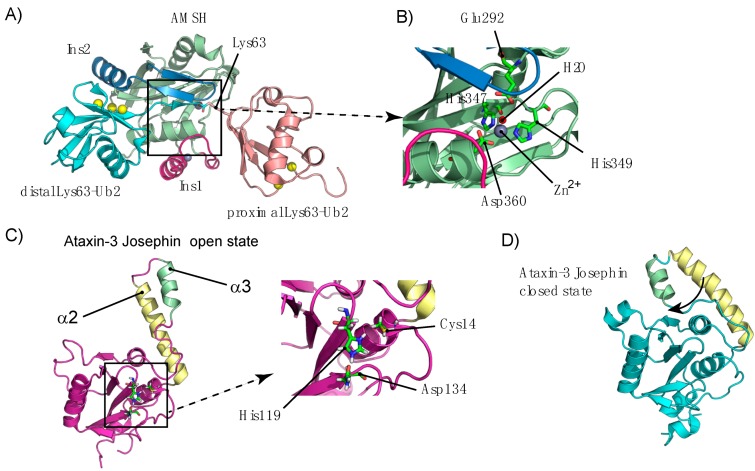
Figure I.
(A) Structure of the AMSH-LP/K63-Ub2 complex (PDB code 2ZNV) showing the JAMM core Ins1 and Ins2. The distal and proximal domains of K63-Ub2 are shown in cyan and pink, respectively, whereas the Ub hydrophobic patch defined by Val70, Ile44 and Leu8 is represented as yellow spheres. (B) The inset represents an expanded view of the catalytic region of AMSH and the coordination of the Zn2+ ion. (C) Structure of the open conformation of the Ataxin-3 Josephin domain (PDB code 2JRI), together with an expanded view of the catalytic triad. (D) Structure of the closed conformation of the Ataxin-3 Josephin domain (PDB code 2AGA).

