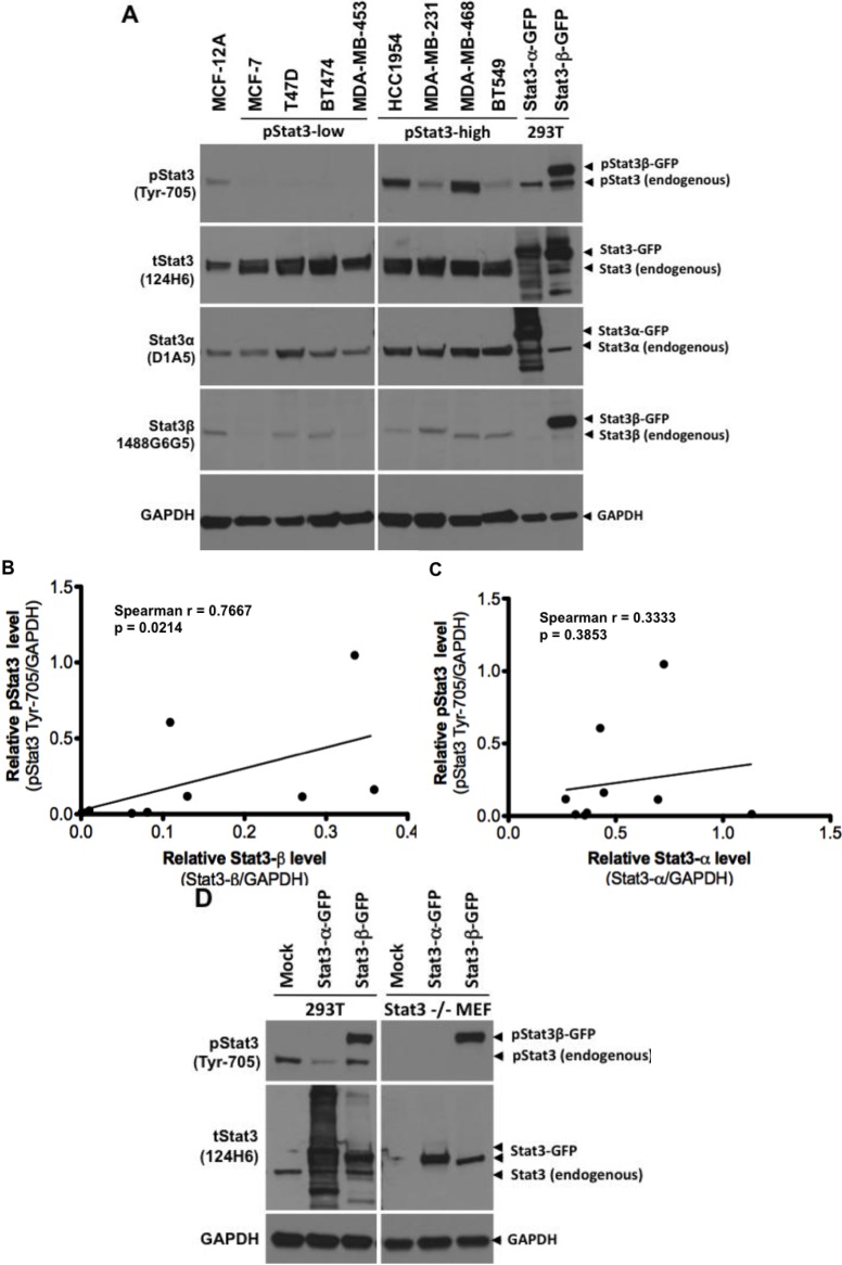
Figure 7.
Stat3β overexpression correlates with Stat3 phosphorylation in breast cancer cell lines. In panel (A), lysates (100 µg protein) from normal breast epithelial cells MCF-12A, and breast cancer cell lines MCF-7, T47D, BT-474, MDA-MB 453, HCC1954, MDA-MB-231, MDA-MB-468, and BT549 cells were subjected to SDS-PAGE, transferred to nitrocellulose membrane and probed for total pStat3 (Tyr-705, clone D3A7), tStat3 (clone 124H6), Stat3α (clone D1A5), Stat3β (1488 G6G5) and GAPDH. Lysates (30 µg of protein) from 293T cells, mock transfected (transfection reagent alone) or transiently transfected with plasmids encoding fusion proteins Stat3α-GFP and Stat3β-GFP were used as controls (lanes 10–11). Stat3β and GAPDH levels were quantified by densitometry and the GAPDH-normalized pStat3 levels plotted as a function of their corresponding Stat3β levels (B, Spearman r = 0.7667, p < 0.05) or Stat3α levels (C, Spearman r = 0.3333, p > 0.05). In panel D, Stat3β-GFP was constitutively phosphorylated when overexpressed in both 293T cells (left panel) and Stat3−/−MEFs (right panel), while Stat3α-GFP was not.

