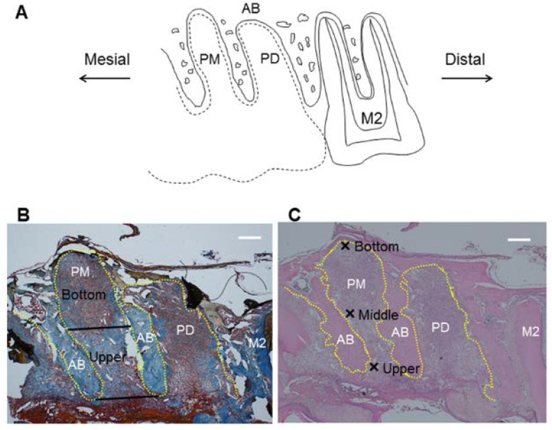
Figure 1.
Measurement regions for histological analysis in the socket after tooth extraction. Sagittal plane of region of interest is shown in (A)—Black dotted line represents extracted maxillary first molar. Regions to evaluate collagen density or FGF-2 expression are shown in (B)—Black straight lines of the alveolar socket represent border of upper area and bottom area. Regions for counting number of polymorphonuclear leukocytes are shown in (C)—Crosses represent upper, middle and bottom parts. Scale bar = 400 μm. Yellow dotted line: outline of alveolar socket. AB: alveolar bone; PM: alveolar socket at palatal mesial root of maxillary first molar; PD: alveolar socket at palatal distal root of maxillary first molar; M2: maxillary second molar.

