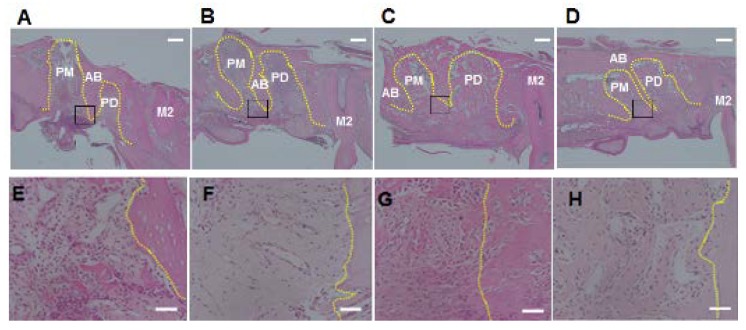
Figure 3.
Photographs of alveolar sockets stained with hematoxylin and eosin at 3 days (A and E: control group; B and F: experimental group) and 8 days (C and G: control group; D and H: experimental group) (Scale bar = 400 μm (A–D) and 50 μm (E–H)). The number of polymorphonuclear leukocytes was low in the experimental group compared with the control group at 3 days. On the other hands, both the groups showed low number of polymorphonuclear leukocytes at 8 days. Yellow dotted line: outline of alveolar socket. Black square: areas of E–H; AB: alveolar bone; PM: alveolar socket at palatal mesial root of maxillary first molar; PD: alveolar socket at palatal distal root of maxillary first molar; M2: maxillary second molar.

