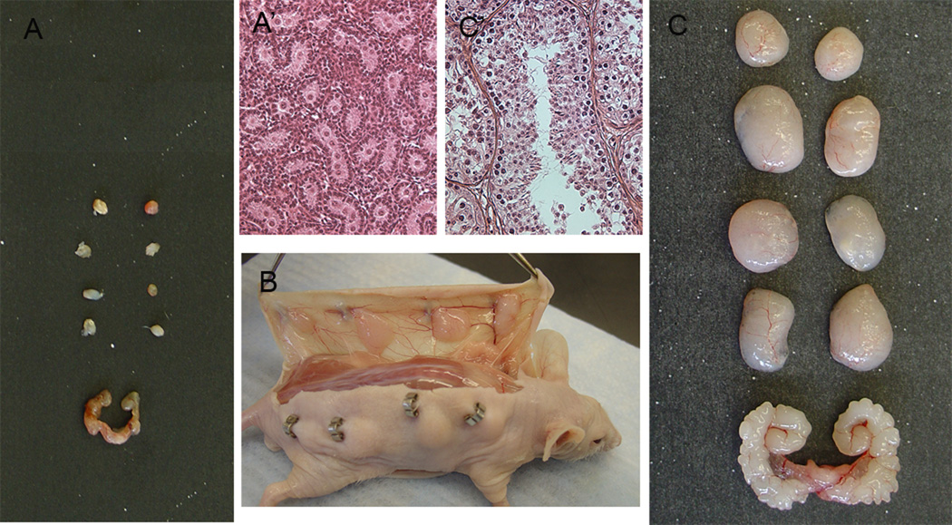
Figure 1.
Xenografting of testis tissue from lambs under the skin of nude mice. A) The size of lamb testis grafts before grafting and seminal vesicle of castrated host mouse and A’) histological appearance of donor tissue showing gonocytes. B) Dorsal and ventral view of the skin of mouse with xenografts 12 weeks postgrafting. C) The size of lamb testis grafts and seminal vesicle of host mouse 16 weeks after grafting and C’) histological appearance of graft tissue showing elongated spermatids. Scale bar, A and C 1cm, A’ and C’ 100µm.

