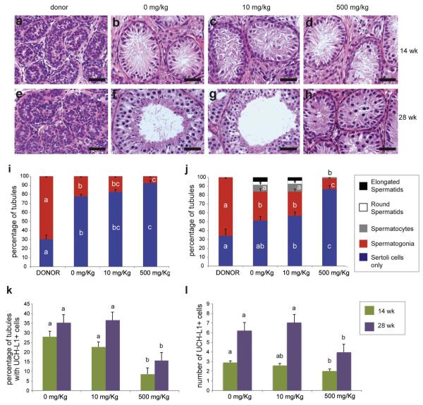
Figure 2.
Development and germ cell differentiation of testis xenografts from mice treated with different doses of DBP. a-h) Histology of donor tissue at the time of grafting (a & e), and that of testis xenografts from mice treated with 0, 10, and 500 mg/kg of DBP for 14 weeks (b-d) and 28 weeks (f-h); i) and j) Percentage of seminiferous tubules with the most advanced type of germ cell present at 14 (i) and 28 (j) weeks; k) Percentage of seminiferous tubules with the presence of spermatogonia (UCH-L1-positive); l) Number of UCH-L1-positive spermatogonia per tubule cross-section. Different letters between bars of the same color indicate statistical difference (P<0.05). Scale bars = 50 μm.

