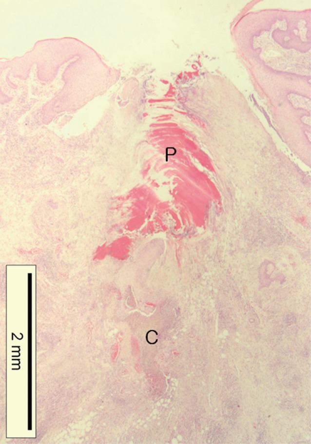
Fig. 4.

Histopathological changes of the tick-feeding lesion; epidermal laceration and invagination, dermal cavitary (C) and perirostral (P) lesion containing abundant amorphous eosinophilic material, and dense inflammatory cell infiltrations around the penetrating site in the dermis and subcutaneous fat tissue (H&E stain, ×12.5).
