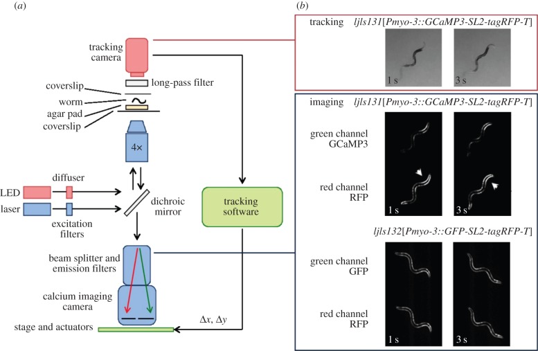
Figure 1.
Tracking calcium imaging microscope. (a) Schematic of the calcium imaging set-up. (b) Image sequences from each camera: worm tracking and re-centring (above) and ratiometric calcium imaging (below). Note increased fluorescence on contracted side of body bends in GCaMP but not control channel (arrowheads).

