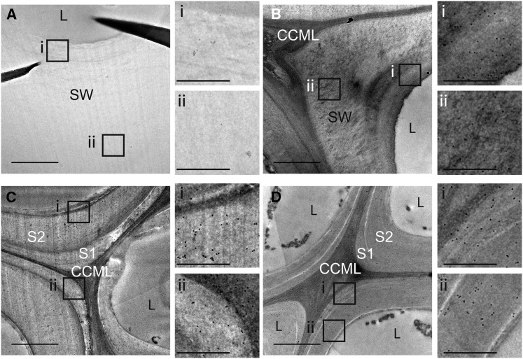
Figure 6.
Immunogold Silver Staining of a Transverse Section of the Flax Stem Median Region with KM1 Antibody.
Bast fibers ([A] and [B]), xylem fibers ([C] and [D]), wild-type ([A] and [C]), and lbf1 ([B] and [D]). CCML, cell corner middle lamella; SW, secondary wall; L, lumen. Smaller photos (i and ii) on the right side of each main photo ([A] to [D]) show a zoom of the corresponding regions indicated on main photo. Bars = 1 μm (main photos) and 0.15 μm (small photos).

