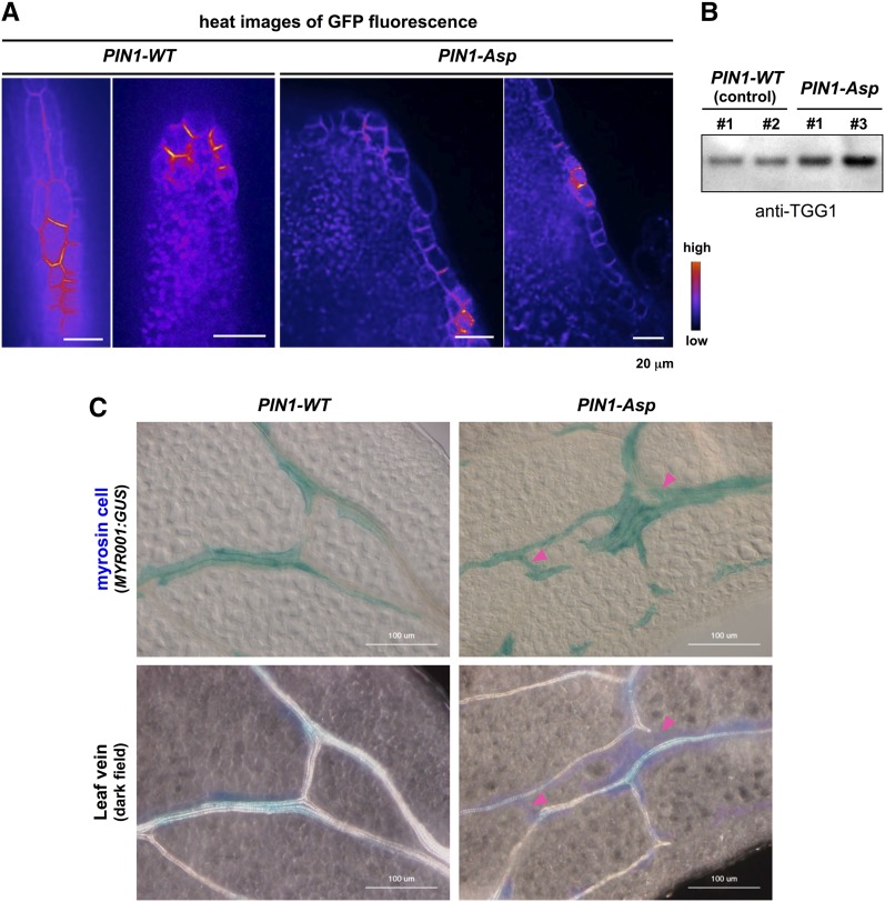
Figure 6.
Expression of Nonpolarizable PIN1 Driven by PIN1 Promoter Phenocopies Abnormal Development of Myrosin Cells.
(A) Subcellular localization of GFP-tagged wild-type PIN1 (PIN1-WT) and nonpolarizable PIN1 (PIN1-Asp) in the leaf margin cells of primordia. Bars = 20 μm.
(B) Rosette leaves expressing PIN1-WT (control) or PIN1-Asp immunoblotted with anti-TGG1 antibody. Numbers (1 and 2 for PIN1-WT; 1 and 3 for PIN1-Asp) indicate independent transgenic lines.
(C) GUS staining (upper panels) and dark-field images (lower panels) of the same areas of MYR001:GUS leaves expressing either PIN1-WT (control) or PIN1-Asp. Arrowheads indicate regions that are positive for myrosin cells and negative for veins. Bars = 100 μm.

