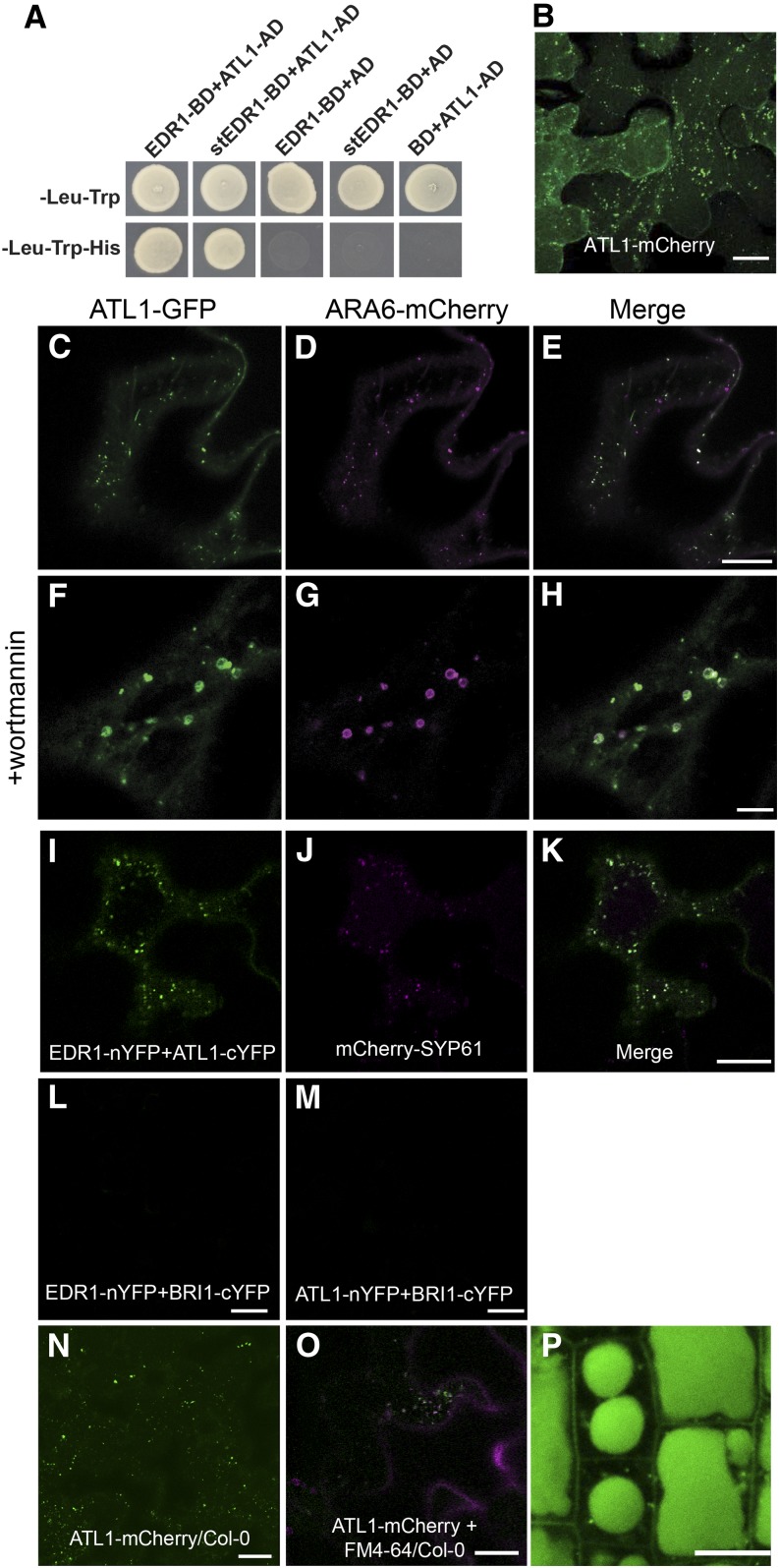
Figure 1.
ATL1 Interacts with EDR1 and Localizes to Endomembrane Structures.
(A) EDR1 and ATL1 interact in a yeast two-hybrid assay. Yeast strains expressing the indicated constructs were plated on double dropout (-Leu-Trp) media to select for the bait and prey plasmids and triple dropout (-Leu-Trp-His) media to select for interaction between the indicated proteins. Empty vectors containing the yeast GAL4 activation domain (AD) or DNA binding domain (BD) were used as negative controls.
(B) ATL1-mCherry localizes to the plasma membrane and intracellular puncta. An N. benthamiana leaf was transiently transformed with a 35S:ATL1-mCherry construct and imaged by confocal microscopy at 40 h after Agrobacterium infiltration. Image is a Z stack composed of eight optical sections.
(C) to (E) ATL1-eGFP colocalizes with the MVB/LE marker ARA6-mCherry. Epidermal cells of N. benthamiana were cotransformed with ATL1-eGFP and the indicated organelle markers fused with mCherry. Cells were imaged using confocal laser scanning microscopy, and single optical sections are shown.
(F) to (H) Leaves from (C) to (E) were infiltrated with 33 μM wortmannin for 1 h to induce MVB dilation.
(I) to (M) EDR1 and ATL1 interact on TGN/EE vesicles in a BiFC assay. EDR1-nYFP and ATL1-cYFP were transiently coexpressed in N. benthamiana with the TGN/EE marker mCherry-SYP61. The BiFC signal is shown in green (I) and the mCherry-SYP61 signal in magenta (J). As negative controls, EDR1-nYFP and BRI1-cYFP were coexpressed (L), as well as BRI1-nYFP and ATL1-cYFP (M).
(N) ATL1-mCherry localizes to the plasma membrane and intracellular puncta in transgenic Arabidopsis. Wild-type Col-0 plants were transformed with a 35S:ATL1-mCherry construct and leaf epidermal cells of 4-week old plants were imaged using CSLM. Image is a Z stack.
(O) ATL1-mCherry colocalizes with the endocytic tracer dye FM4-64 in stable transgenic Arabidopsis lines. Image was taken 15 min poststaining.
(P) ATL1-mCherry accumulates inside the lytic vacuoles of root epidermal cells.
Bars = 10 μm.

