Figure 6.
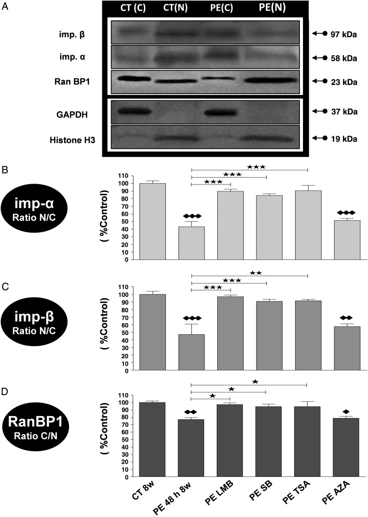
Alteration in the expression of cytosolic and nuclear transport receptors (Imp-α and Imp-β) and transport driving force protein (RanBP1) in PE-treated adult rat CM through p38 MAPK, HDAC CL II, and CRM1, but not GSK-3β pathway activation. Representative western blots (A) and analysis by densitometry of Imp-α (B), Imp-β (C), and RanBP1 (D) expressions in the nuclear and cytosolic fractions of rat control CM (CT 8w) exposed to PE (PE 48 h 8w) in the absence or presence of SB, TSA, LMB, and AZA. Even sample loading of nuclear fractions was confirmed by Histone H3 detection and cytosolic fractions by GAPDH detection. As for equal sample loading between nuclear and cytosolic fractions together, this was performed by staining the blots with Coomassie blue. Results are expressed in percent control and are shown as mean ± SEM (n = 3); * and ♦P < 0.05, ** and ♦♦P < 0.01, *** and ♦♦♦P < 0.001; ♦ or ♦♦ or ♦♦♦ are vs CT8w.

