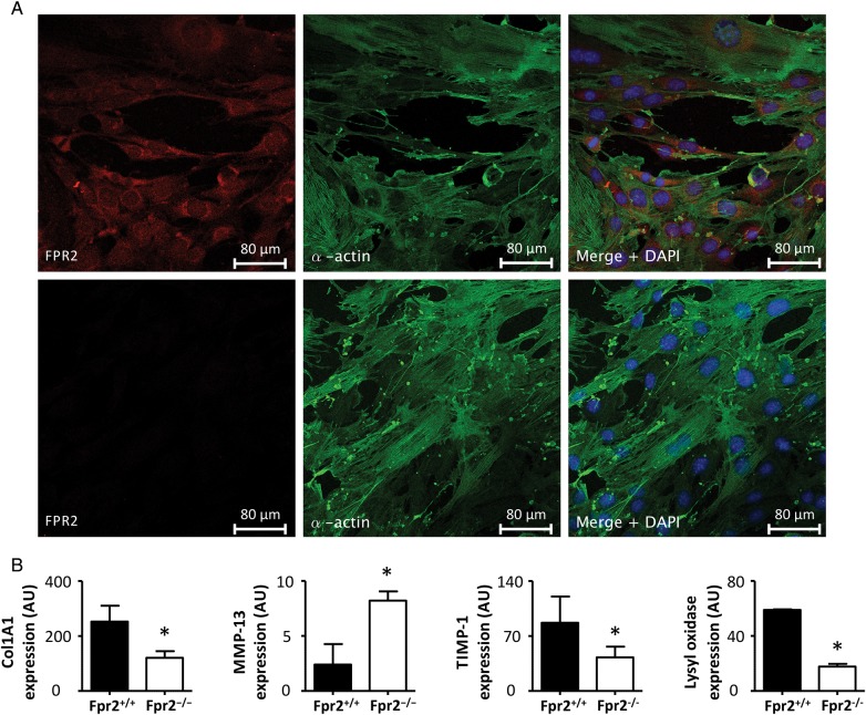
Figure 6.
(A) Representative confocal IF stainings of FPR2 (red) and α-smooth muscle actin (green) in mSMC derived from either Fpr2+/+ (top) or Fpr2−/− mice (bottom). (B) The results of real-time PCR for collagen 1A (Col1A1), lysyl oxidase (LOX), MMP–13, and tissue inhibitor of MMP (TIMP)-1 in mSMC derived from Fpr2+/+ and Fpr2−/− mice (B; N = 3). *P < 0.05 compared with the respective control in each graph.

