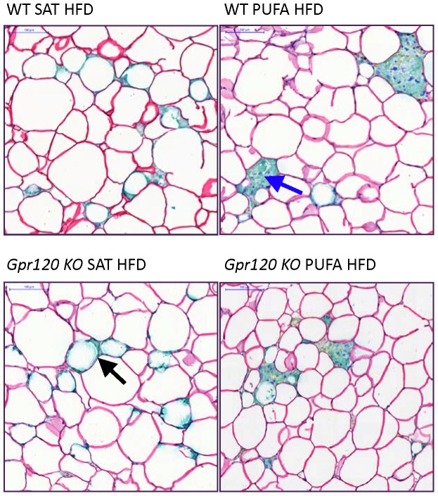
Figure 6. Adipose tissue histology.
Representative slides of epididymal WAT double-stained for Perilipin and Mac2 (Macrophage 2 antigen, Galectin-3) from WT and Gpr120 KO mice fed either the SAT HFD or PUFA HFD as indicated. Perilipin staining is seen as read coloured lines surrounding the cells. Some cells, typically associated with ‘crown like’ structures (CLS) do not display perilipin staining. Arrows indicate CLS (black arrow) and multinuclear giant cell aggregate (blue arrow). Scale bar upper left corner = 100 µm.

