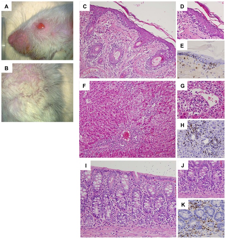
Figure 3. Acute GVHD in the skin, liver, and intestine after allogeneic bone marrow transplantation (BMT).
On day 28 after allogeneic BMT, macroscopic findings of the skin indicated erythematous rush and alopecia (A, B). Light microscopic findings showed inflammatory cells, mainly CD3+ T-cells, infiltrating the epidermis and the hair follicle in the dermis, indicating acute GVHD in the skin (C, D: HE stain, E: CD3 stain, C: ×400, D, E: ×800). Inflammatory cells, mainly CD3+ T-cells, infiltrated the portal areas and spread to the hepatic lobules with cholangiolitis and phlebitis in the portal and central veins, indicating acute GVHD in the liver (F, G: HE stain, H: CD3 stain, F: ×400, G, H: ×800). In the colon, erosion and inflammatory cell infiltration was noted with cryptitis and infiltration of CD3+ T-cells, indicating acute GVHD in the colon (I, J: HE stain, K: CD3 stain, I: ×600, J, K: ×800).

