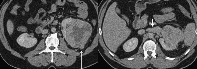
Figure 6.

A 42-year-old female who underwent total left nephrectomy for a grade 3, clear-cell renal cancer. Macroscopic venous, perinephric fat and sinus fat invasions were found. These two axial contrast-enhanced CT images demonstrate left renal vein invasion (short arrow) and nodular breach of the renal capsule (long arrow). The images show tumour necrosis, increased stranding of the perinephric fat, thickening of the perirenal fascia and tumour edge abutting the perirenal fascia and sinus structures, and all these features are associated with an increased likelihood of Stage T3a disease.
