
Figure 3.
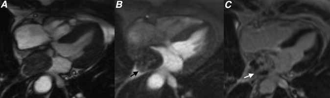
(a) Cine steady-state free procession, (b) first pass perfusion and (c) late gadolinium enhancement (LGE) four-chamber sequences showing a left atrial myxoma. Note how the lesion shows limited enhancement on first pass perfusion sequences (black arrow) and heterogeneous enhancement on LGE sequences (white arrow).