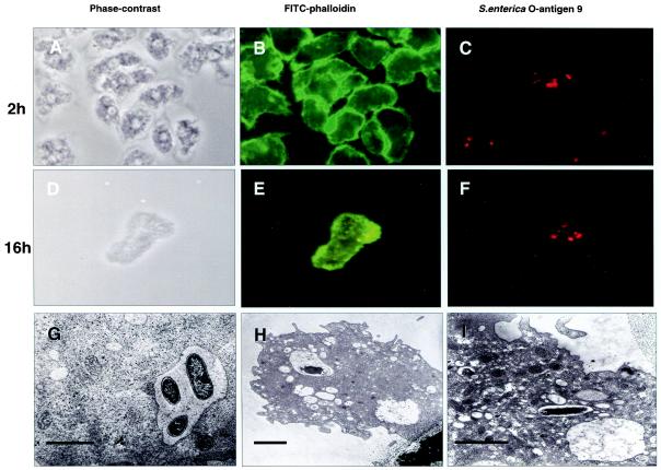
FIG. 6.
(A through F) Microscopy of A. rhysodes infected with invasive Salmonella serovar Dublin at 2 h (A through C) and 16 h (D through F) postinfection. (A and D) Phase-contrast images; (B and E) staining of cells with fluorescein isothiocyanate (FITC)-phalloidin; (C and F) staining of bacteria with a rabbit antiserum against S. enterica O-antigen 9 and rhodamine-conjugated anti-rabbit immunoglobulin G. (G through I) Transmission electron microscopy shows intravacuolar Salmonella serovar Typhimurium bacteria at 2 h postinfection (G) and Salmonella serovar Dublin at 16 h postinfection (H and I). (G) Magnification, ×5,000; bar, 3.4 μm. At this magnification, bacterial membranes appear intact. (H) Magnification, ×5,000; bar, 2.2 μm. (I) Magnification, ×8,000; bar, 2.3 μm.

