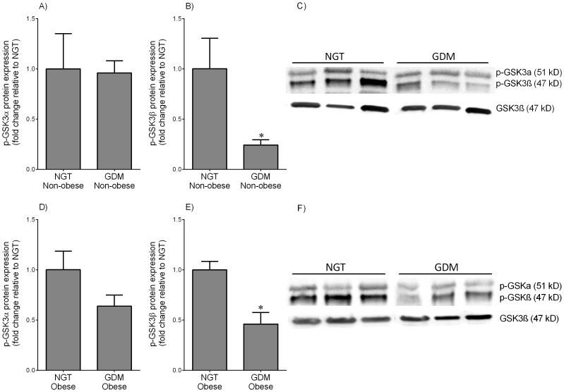
Figure 2. Phosphorylated GSKα/β expression in skeletal muscle from NGT and GDM women.
Skeletal muscle was obtained from (A–C) non-obese and (D–F) obese women with NGT (n = 6 patients per group) and diet-controlled GDM (n = 6 patients per group) at the time of term Caesarean section. Phosphorylation of GSK3α at serine 21 (p-GSKα) and GSK3β at serine 9 (p-GSKβ) was analysed by immunoblotting and normalised to total GSK3β protein expression. The fold change was calculated relative to NGT and data is displayed as mean ±SEM. *P<0.05 vs. NGT (Student's t-test). Representative Western blot from 3 NGT and 3 diet-controlled GDM patients is also shown.

