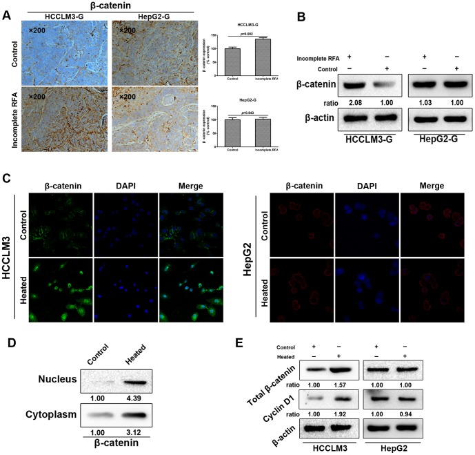
Figure 5. Incomplete RFA induced overexpression or internalization of β-catenin accompanied with EMT in vivo and in vitro of HCCLM3 cells.
(A) Immunohistochemistry revealed that β-catenin expression was increased in incomplete RFA group of HCCLM3-G xenografts (P = 0.002); in HepG2-G xenografts, there was no significant difference between two groups in β-catenin expression (P = 0.843). (B) Western blot showed the enhanced β-catenin expression in the tumor tissues after incomplete RFA compared with the controls of HCCLM3-G xenografts. (C) Immunofluorescence staining showed the strong cytoplasmic and nuclear localization of β-catenin in heat-treated HCCLM3 cells, and the typical membranous β-catenin expression in the cell-cell contacts of HCCLM3 cells in control. However, there was no significant difference between two groups in β-catenin expression of HepG2 cells. (D) Distribution of β-catenin in cytoplasm and nucleus of HCCLM3 cells was detected by western blot in vitro. (E) Western blot analysis exhibited the expression of total β-catenin and its downstream target gene Cyclin-D1 of both two HCC cells in vitro.

