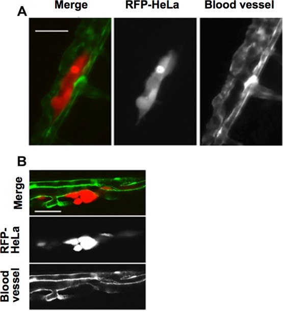
Figure 1. Extravasation of cancer cells after severe emboli formation in arterioles of zebrafish larvae.

(A) RFP-HeLa cells and endothelial cells in arterioles were examined under an epifluorescence microscope 17–20 h after formation of the emboli. (B) The spatial locations of the extravasated RFP-HeLa cells and blood vessels were examined under a confocal microscope. Bars, 100 µm (A) or 40 µm (B).
