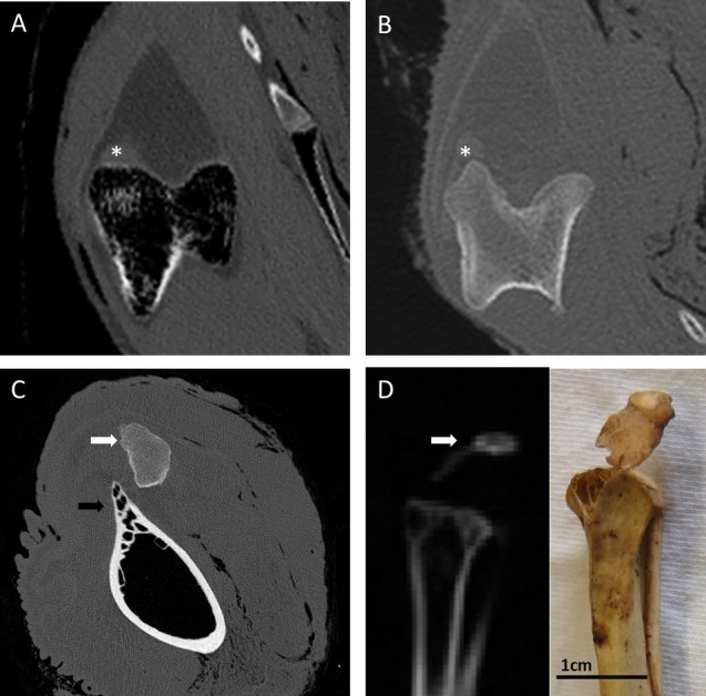
Figure 3. CT images of the patellar region in select Palaeognathae.

(A) Emu (Dromaius novaehollandiae) distal femur in transverse section, with the lateral femoral condyle on the left. There is no mineralised patellar structure, and the patellar tendon itself is radiolucent with regard to surrounding soft tissues. The soft tissue opacity near the lateral condyle (∗) represents the m. tibialis cranialis muscle origin. (B) Southern cassowary (Casuarius casuarius) distal femur in transverse section, with the lateral femoral condyle on the left. There is no mineralised patellar structure, and like the emu, the patellar tendon itself is slightly radiolucent with regard to surrounding soft tissues. The soft tissue opacity near the lateral condyle (∗) represents the m. tibialis cranialis muscle origin. (C) Ostrich (Struthio camelus) distal femur in transverse cross section, with bony outcrop which becomes the lateral femoral condyle on the left (black arrow). The ossified proximal patella (white arrow) overlies the region of the lateral condyle and patellar sulcus. (D) Red-winged tinamou (Rhynchotus rufescens specimen NHMUK 1851.11.10.43) proximal tibiotarsus in sagittal cross-section, with the mineralised patella (white arrow) attached by a slip of desiccated patellar tendon, as can be seen grossly (photo insert).
