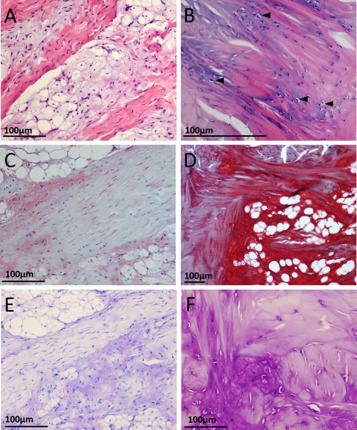
Figure 6. Appearance of cartilage-like tissue within the emu patellar tendon at five weeks (left) and 18 months (right) with different histological stains.
(A) Five weeks with H&E. The tissue is basophilic and vesicular in appearance, often seen in association with collagen fibre bundles. (B) 18 months with H&E, from a region near the proximal muscle junction. The tissue has a more homogenous and less vesicular appearance. Chondrocyte-like cells (filled arrows) sit within the normally eosinophilic collagen fibre bundles, creating an intense localised basophilia. (C) Five weeks with Safranin O/fast green. (D) 18 months with Safranin O/fast green. (E) Five weeks with toluidine blue. (F) 18 months with toluidine blue. Nearby collagen fibres bundles near the chondrocyte-like cells show some metachromatic (purple) staining.

