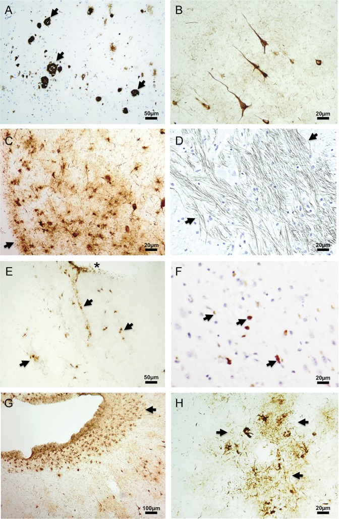
Figure. Immunohistochemistry staining of brain sections from chronic traumatic encephalopathy case.
A 9-µm paraffin section from (A) temporal cortex with focus of β-amyloid-positive neuritic plaques (black arrows), (D) medulla with tau immunoreactivity in neuronal processes (black arrows), and (F) hippocampus with TDP-43 aggregates (black arrows). Fifty-micrometer fixed free-floating sections from (B) frontal cortex with cortical layer I–II tau-positive neurofibrillary tangles, (C) pons with tau-immunoreactive glia (black arrow pointing to pial surface), (E) spinal cord with tau immunoreactivity glia (black arrows; *central canal), (G) temporal cortex with subpial tau immunoreactivity in depth of sulcus (black arrow), and (H) frontal cortex with tau immunoreactivity surrounding a blood vessel (black arrows). Antibodies: (A) monoclonal β-amyloid 4G8, (B–C, E, G–H) monoclonal p-tau AT8, (D) monoclonal p-tau PHF1, (F) polyclonal p-TDP43. Representative sections are shown. Section sampling for the entire case (exceeding National Institute on Aging–Alzheimer's Association guidelines) included 28 sections from 8 different regions of neocortex, hippocampus, amygdala, entorhinal cortex, 2 levels of basal ganglia, hypothalamus, thalamus, midbrain, pons, medulla, cerebellum, all levels of spinal cord, and dorsal root ganglia.

