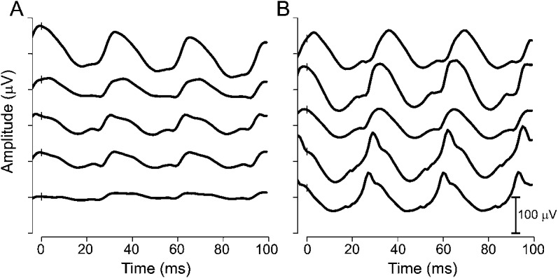
Figure 1. Raw ERG waveforms from a child who developed VGB-RD and one who did not.

Light-adapted 2.29 flicker waveforms plotted against time (see insert for amplitude scale). (A) Male diagnosed with IS at 12 months of age and started VGB at 13 months of age. ERG waveforms are shown for the baseline test (top trace) and for 4 subsequent assessments at 3, 6, 10, and 13 months of VGB treatment. VGB-RD occurred after 3 months of VGB treatment. (B) Male diagnosed with IS at 4 months of age and started VGB at 7 months of age. ERG waveforms are shown for the baseline test (top trace) and for 4 follow-up assessments at 2, 6, 12, and 16 months of VGB treatment. There was no evidence of VGB-RD. ERG = electroretinogram; IS = infantile spasms; VGB = vigabatrin; VGB-RD = VGB-induced retinal damage.
