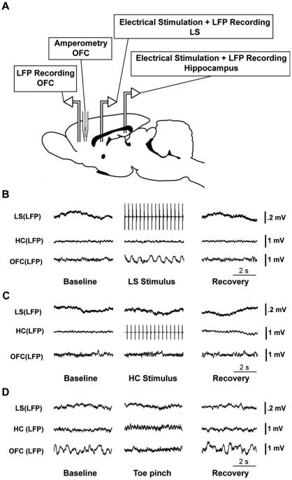
Figure 1.
Recording setup and examples of delta power changes in OFC. (A) Schematic diagram of animal preparation for experiments. Bipolar electrodes for stimulating and/or recording local field potential (LFP) were placed in hippocampus (HC) and in the orbital frontal cortex (OFC) on the left side, as well as in the lateral septum (LS) on the right side (left and right sides are not indicated on the diagram). In addition, a choline microelectrode was implanted into the OFC on the right side (homologous coordinates to left OFC LFP electrodes). (B) An example of LFP recordings during lateral septal stimulation under light anesthesia. LFP signals from LS, HC and OFC are shown. Electrical stimulation of LS at 3 Hz causes slow oscillations in the OFC, followed by return to fast activity during the recovery period. (C) An example of LFP recordings during hippocampal stimulation under light anesthesia. LFP signals from LS, HC and OFC are presented. Electrical stimulation of HC at 3 Hz causes no obvious changes in either LS or OFC. (D) Example of LFP recordings in LS, HC and OFC during toe pinch. Animal is under deep anesthesia with slow wave activity in OFC at baseline. Toe pinch causes a decrease in slow wave activity in OFC which then returns during recovery.

