
Figure 2.
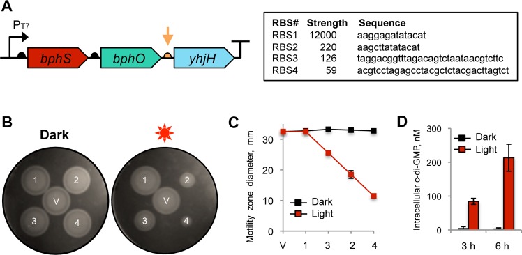
Synthetic operon for light-activated c-di-GMP synthesis. (A) Structure of the synthetic operon for light-activated c-di-GMP synthesis. The genes encoding light-activated DGC (bphS), heme oxygenase (bphO), and c-di-GMP PDE (yhjH) are assembled in a single operon, bphS-bphO-yhjH. A semicircle in front of each gene indicates a RBS; a T-sign at the end of the operon indicates a transcription terminator. The expression levels of yhjH were altered by using RBS sequences (orange arrow) of varying strengths shown in the box. (B) Adjustment of the yhjH RBS strength using semisolid agar motility assays in E. coli MG1655[DE3]. V, pMQ56 (empty vector); 1, RBS1; 2, RBS2; 3, RBS3; 4, RBS4. Increased intracellular c-di-GMP levels decrease the size of a motility zone. (C) Diameters of the swimming zones from panel B. (D) Intracellular c-di-GMP levels measured in liquid-grown cultures of MG1655[DE3] expressing bphS-bphO-yhjH with RBS3 upstream of yhjH.