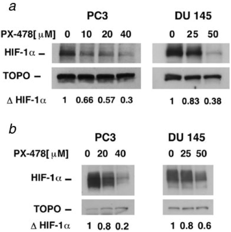
Figure 1.

Western blot analysis of HIF-1α in PC3 and DU 145 cells. (a) Cells were treated with PX-478 for 20 hr under normoxic condition. (b) Cells were treated with PX-478 for 20 hr under normoxic condition, exposed to 1-hr hypoxic gassing and analyzed. ΔHIF-1α: fold change in HIF-1α compared to the control. For normoxic samples, 60 μg protein, and for hypoxic samples, 15 μg protein was analyzed.
