
Figure 6.
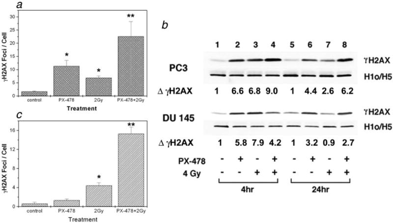
Analysis of γH2AX following treatment with PX-478 and radiation. (a) γH2AX foci per cell in PC3 cells. Cells were treated with 20 μmol/l PX-478 for 18 hr, irradiated and after 1 hr changed to drug-free media. Cells were incubated for another 24 hr in drug-free media and γH2AX foci were counted in ~70 cells. Each data point represents Av ± SEM of 3 separate experiments. *Compared to the control, **compared to the 2Gy. (b) Western blot analysis of γH2AX in PC3 (top) and DU 145 (bottom) cells. Cells were treated as above and γH2AX was analyzed at 4 hr and 24 hr after radiation. Histone H1o/H5 was used as a loading control. ΔγH2AX: fold change compared to the control. Data shown are representative of 3 separate experiments. (c) Effect of short exposure to PX-478 on H2AX phosphorylation in PC3 cells. Cells were treated with PX-478 for 30min and changed to drug-free media. After 1h cells were irradiated and H2AX foci/cell were counted at 4h (data not shown) and at 24h. *Compared to the control, **compared to the 2Gy.