Figure 1.

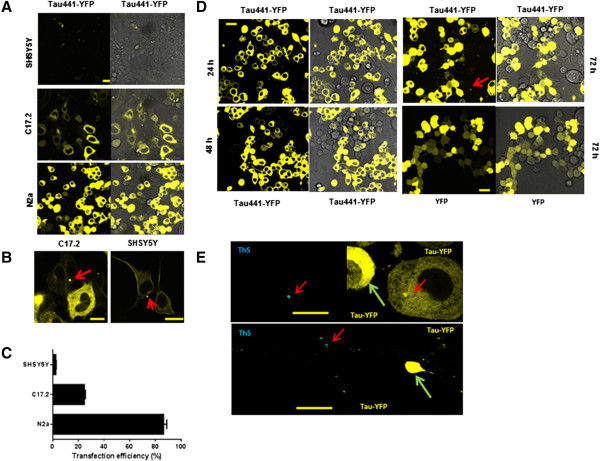
Expression and aggregation of Tau441-YFP in three different cell types. A) SHSY5Y, C17.2 and N2a cells were transfected with Tau441-YFP and live cell images were taken at 24 h with a confocal microscope. Darkfield images showed better fluorescence signal and phase contrast provided images for the whole cell. B) Aggregates formed in SHSY5Y and C17.2 cells (red arrow). C) Quantification of transfection efficiency was done with Image J. D) Time course of Tau441-YFP expression and aggregation in N2a cells. Aggregates appeared at 72 h after transfection while no aggregates formed in YFP transfected cells, which suggested that those aggregates were tau specific. E) 72 h after transfection, cells were fixed with 4% PFA and stained with thioflavin-S (ThS). Only a small population of cells showed ThS-positive aggregates (red arrow). Cells (green arrow) with diffusely distributed tau were ThS negative even though a high level of Tau441-YFP was expressed in those cells. This further confirmed that the ThS signal was not a false signal due to the leakage of YFP. Magnification: 63x. Scale bar: 10 μm.
