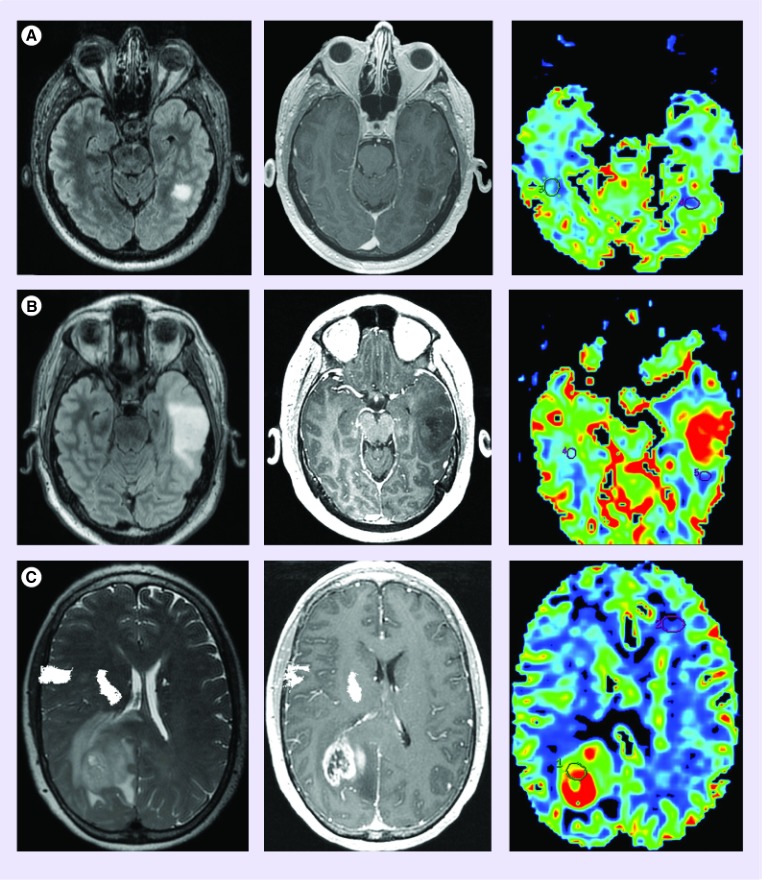
Figure 4. . Cerebral blood volume differentiates high from low-grade glioma.
(A) Diffuse astrocytoma (WHO grade II) is morphologically manifested as a fluid-attenuated inversion recovery hyperintense (FLAIR; left) nonenhancing (middle) mass with cerebral blood volume (right) measurements similar to normal appearing white matter. (B) Anaplastic astrocytoma (WHO grade 3) typically presents as a FLAIR hyperintense mass with minimal if any contrast enhancement; however, unlike low-grade glioma, demonstrates elevated cerebral blood volume. The presence of increased perfusion metrics within a nonenhancing glioma suggests the presence of aggressive biological features that portend a high-grade diagnosis. (C) glioblastoma (WHO grade 4) can present as a rim enhancing T2/FLAIR hyperintense mass with markedly elevated cerebral blood volume.

