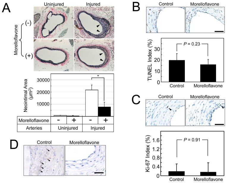
Fig. 4. Morelloflavone inhibits injury-induced neointimal proliferation in a mouse carotid artery injury model.
(A) Verhoeff–van Gieson (VVG) staining of mouse carotid arteries. Upper panel: Photomicrograms of the carotid arteries. Uninjured, right carotid arteries that are sham operated; injured, left carotid arteries in which endothelial cells were denuded by the insertion of an epoxy resin probe; Arrows, neointimal formation. Lower panel: Morphometric analyses of injured and uninjured mouse carotid arteries. *, P < 0.01 (Two-sample t-test; n = 9 for control; n = 10 for morelloflavone-treated). Morelloflavone significantly blocked injury-induced neointimal formation in mouse carotid arteries. (B) TUNEL apoptosis assays. The TUNEL indices were determined as the number of cells with TUNEL-positive nuclei divided by the total number of cells counted and expressed as a percentage (n = 9 for control; n = 10 for morelloflavone-treated). Size bar = 25 μm. (C) Ki-67 proliferation assays. The Ki-67 indices were determined as the number of cells with Ki-67-positive nuclei divided by the total number of cells counted and expressed as a percentage. Size bar = 25 μm. Oral morelloflavone treatment resulted in reduced neointimal formation, without increasing apoptotic or proliferating cells in the neointima. (D) p-ERK staining. Phosphorylated ERK was detected by using anti-p-ERK antibody. Size bar = 25 μm. The nuclear p-ERK signals (black arrows) were seen in 42.9 % of the neointima of control animals and in 12.5 % of the neointima of morelloflavone-treated animals (n = 7 and 8, respectively). Oral morelloflavone treatment was associated with reduced p-ERK positivity in the neointima.

