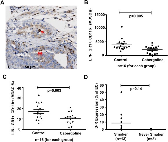
Figure 6.

Tumor infiltrating myeloid derived suppressor cells decreased in vivo by D2R agonists and endothelial D2R expression increased in NSCLC patients with a smoking history. A: Representative image of D2R immunohistochemistry of human lung cancer tissue microarray depicting D2R‐positive myeloid/macrophage lineage cells (red arrows). B–C: C57BL/6 wildtype mice were orthotopically injected with 1 × 10⁵ LLC1 cells. Mice were intraperitoneally administered vehicle or cabergoline daily for seven days starting on day five post‐injection of LLC1 cells. After 12 days, the lungs were harvested from euthanized mice and cells were extracted through collagenase digestion. Flow cytometry was used to determine the number (B) and percentage (C) of murine myeloid derived suppressor cells (MDSCs), which were defined as LIN‐, GR1+, and CD11b+. n = 16 mice/treatment group. D: Immunohistochemistry was performed using a monoclonal D2R antibody on a human lung cancer tissue microarray. The graph shows the percentage of endothelial cells stained positive for D2R among lung cancer tissue obtained from smokers (n = 13) compared to those from never smokers (n = 3).
