
Figure 1.
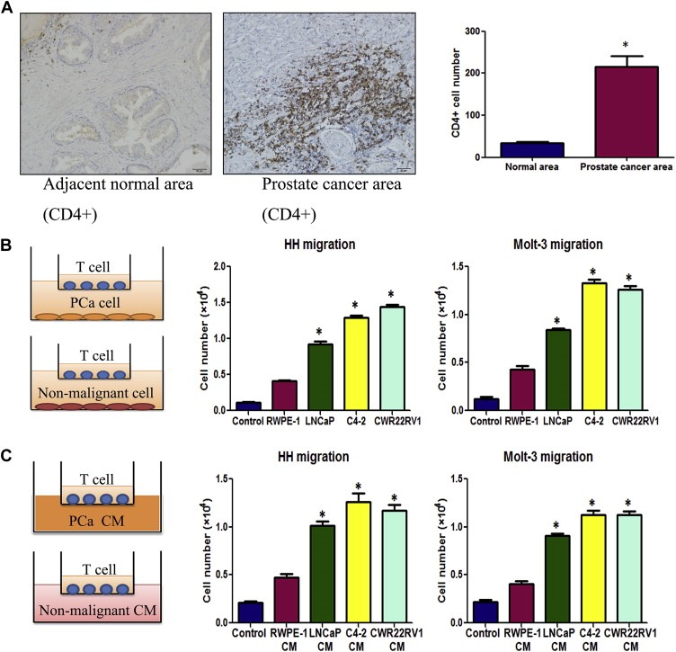
Prostate cancer tissues recruit more T cells. A. Clinical specimen IHC staining from 20 prostate cancer patients showed that more CD4(+) T cells infiltrate surrounding the prostate cancer area than in the adjacent normal prostate area, scale bar: 20 μm, *P < 0.05. B. Transwell T cell migration assay. 1 × 10⁵ of PCa cells or non‐malignant prostate epithelial cells were plated into the lower chambers of the transwells. 1 × 10⁵ of CD4(+) T cells, HH cells or Molt‐3 cells, were plated onto the upper chamber with 5 μm pore polycarbonate membrane for T cell migration assay. The cells migrated into the lower chamber media were collected after 6 h and counted by the Bio‐Rad TC10 automatic cell counter. Compared to the prostate normal epithelial cell line RWPE‐1, LNCaP, C4‐2 and CWR22RV1 recruited more HH and Molt‐3 (*P < 0.05). C. When PCa or RWPE‐1 conditioned media (CM) was added into the lower chamber, PCa cell CM could attract more HH or Molt‐3 cells compared with RWPE‐1 cells CM (*P < 0.05).