
Figure 6.
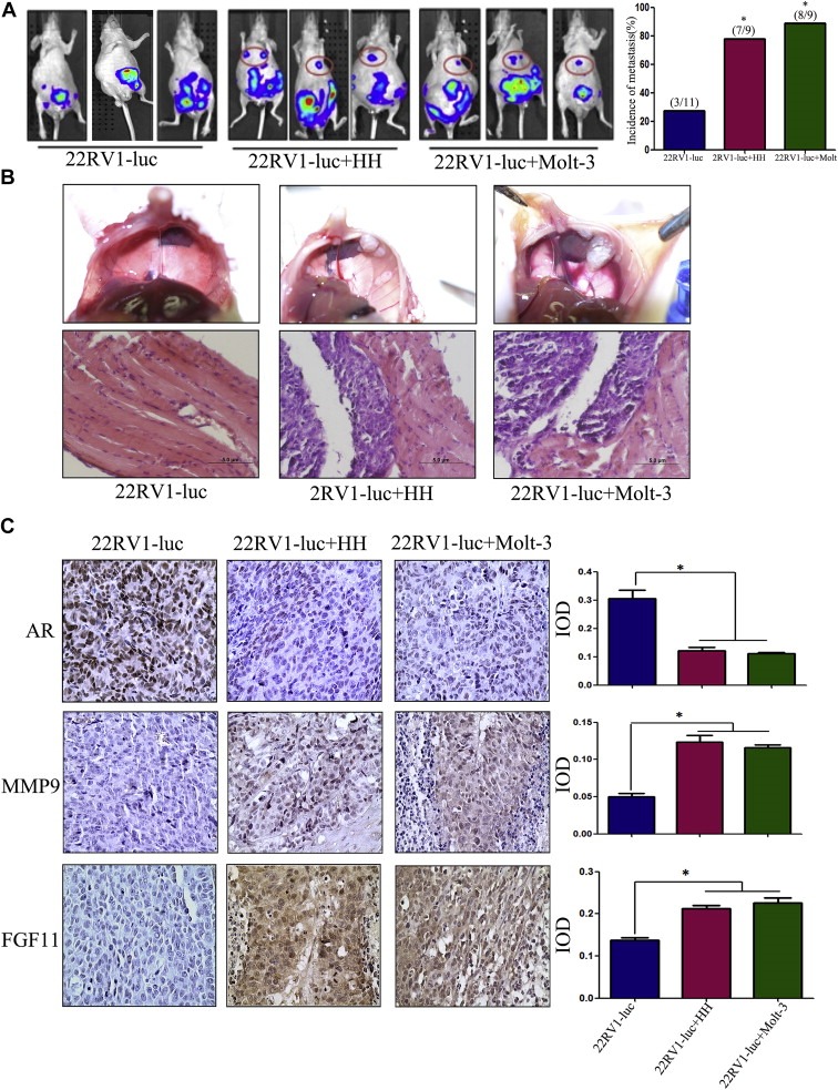
Induction of PCa metastasis by T cells in orthotopic PCa model. A. CWR22RV1‐luc (22RV1‐luc) cells (1 × 10⁶) were mixed with HH or Molt‐3 (1 × 10⁵) and orthotopically implanted into the anterior prostates of nude mice. After 6 weeks, the PCa growth and metastases were evaluated by the IVIS system. The 22RV1 cells co‐injected with T cells showed significant increase of the distant metastatic tumors in diaphragm, compared to the 22RV1 single injection mice (*P < 0.05). B. The mice were then sacrificed (6 week after implantation), the metastatic tumors observed on the diaphragm were confirmed by histology H&E staining. C. IHC staining confirmed T cells co‐implanted 22RV1 tumors have a reduced AR, increased MMP9 and FGF11 expression (400×) (*P < 0.05).