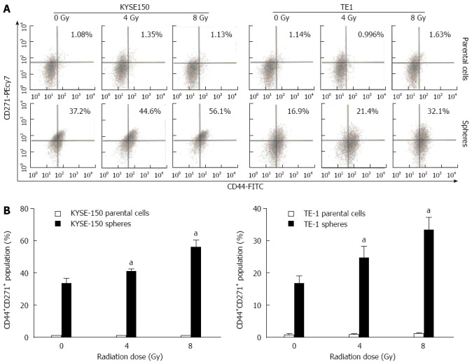
Figure 4.

Proportion changes of CD44+CD271+ cells in parental cells and stem-like spheres with different doses of radiation. A: Expression of CD44+, CD271+ and CD44+CD271+ were detected by flow cytometry; B: The differences in CD44+CD271+ cell proportion in parent cells and cell spheres under different irradiation doses were significant. In addition, the proportion of KYSE150 and TE1 cell spheres under irradiation doses of 4 and 8 Gy was significantly different from that of 0 Gy, aP < 0.05 vs control group.
