
Figure 1.
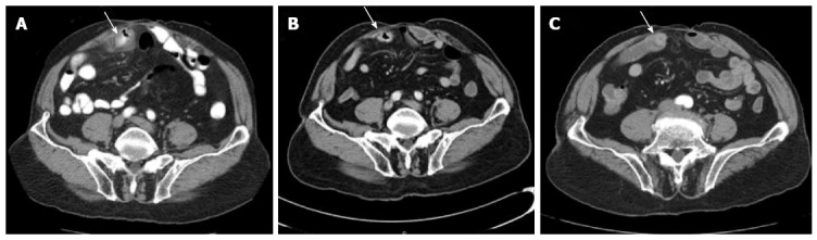
Selected computed tomography images showing clinical course of perforated small bowel diverticulum. A: Computed tomography (CT) scan at time of initial presentation showing a focally thickened loop of small bowel with a small collection adjacent to the thickened small bowel concerning for a perforated small bowel diverticulum; B: Repeat CT scans 9 d later depicting resolution of the inflammatory changes but persistent thickening; C: CT enterography 40 d after admission after complete treatment course of antibiotics showing resolution of all inflammatory changes and imaging consistent with a small bowel diverticulum. The arrow is pointing out the small bowel diverticulum and its evolution over the next few months.