
Figure 2.
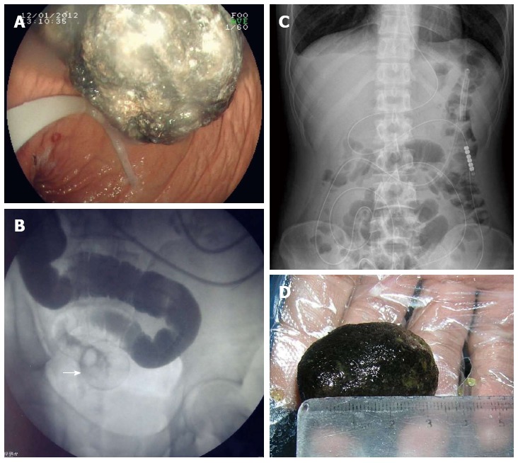
Treatment procedures. A: A greenish, semisolid mass in the stomach observed on gastroscopy; B: Imaging revealed slight expansion of the intestine and dislodgement at a round point (white arrow); C: No air-fluid level was observed on plain abdominal radiograph two days later; D: Image of the discharged diospyrobezoar.