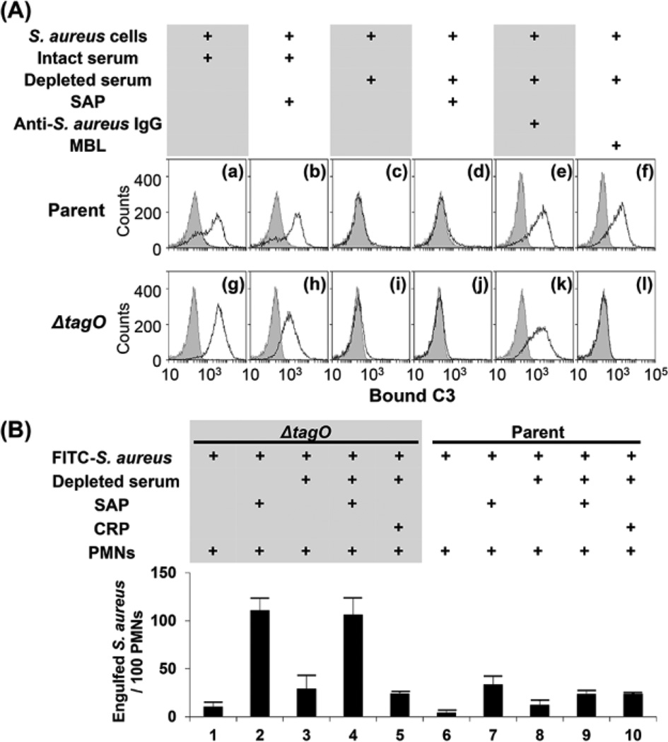
Figure 4. SAP does not induce C3 deposition on Δspa mutant or ΔtagO, Δspa double mutant cells, but induces complement-independent phagocytosis of S. aureus cells by PMNs.
(A) Panels (a) and (g), C3 deposition on parental and ΔtagO mutant cells after incubation with intact serum; (b) and (h), C3 deposition by incubation of SAP (1 µg) with intact serum; (c) and (i), C3 deposition in depleted serum; (d) and (j), C3 deposition after addition of 1 µg SAP into depleted serum; (e) and (k), C3 deposition after addition of S. aureus-recognizing Ig (1 µg) into depleted serum; (f) and (l), 50 µg MBL added into panels (c) and (i), respectively. The serum concentration used was 10%. C3b bound to S. aureus cells was detected by flow cytometry as described in Materials and Methods. The curves with gray areas underneath represent cell-only controls. (B) Ethanol-killed ΔtagO mutant (columns 1–5) and parental cells (columns 6–10) were labeled with FITC (0.1 mM) and incubated with depleted serum (10%) in a 20 µl buffer. SAP (1 µg) or CRP (1 µg) and FITC-labeled S. aureus cells (3.8 × 10⁶ cells) were incubated with human PMNs (1.5 × 10⁵ PMNs) at multiplicity of infection of 25 in 40 ml RPMI 1640 medium at 37°C for 1 h. Phagocytosed S. aureus cells in 100 PMNs were counted under fluorescent phase-contrast microscopy. Data are the means ± SD of results of three independent experiments. *p < 0.01.

